PLAQUE INDUCED GINGIVITIS
Plaque-induced gingivitis is inflammation of the gingiva resulting from bacteria located at the gingival margin. Gingivitis is prevalent at all ages of dentate population and is considered to the most common form of periodontal disease.
Loe (1965) performed a study on gingivitis where 12 healthy individuals with clinically normal gingiva and good oral hygiene withdrew all forms of oral hygiene. This resulted in accumulation of soft debris and marginal gingivitis in all subjects. The time necessary to develop gingivitis varied from 10 to 21 days. Some had gingivitis after 10 days, but majority of the subjects required 15-21 days. Reinstitution of oral hygiene resulted in healthy gingival conditions and re-establishment of the original bacterial flora within 5-10 days.
Page and Schroeder (1976) demonstrated that initial changes from health to plaque-induced gingivitis are not detectable clinically.
DENTAL PLAQUE
- An organized mass, consisting mainly of microorganisms
- Host associated biofilm
-
May be readily visualized on teeth after 1 to 2 days with no oral hygiene measures

CHARACTERISTICS OF PLAQUE INDUCED GINGIVITIS
- Plaque present at gingival margin
- Disease begins at the gingival margin
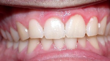
- Change in gingival colour
- Change in gingival contour
- Sulcular temperature change
- Increased gingival exudate
- Bleeding upon provocation
- Absence of attachment loss
- Absence of bone loss
- Histological changes
- Reversible with plaque removal
DEVELOPMENT OF GINGIVITIS
INITIAL LESION
The initial lesion appears 2-4 days after plaque accumulation in previously healthy gingiva and is localized to the gingival sulcus. The initial inflammatory change occurs in response to microbial activation of resident leukocytes and the subsequent stimulation of endothelial cells. This lesion is not clinically apparent.
- Characteristics
- Vascular dilation and vasculitis subsequent to the junctional epithelium
- Infiltration of polymorphonuclear neutrophils (PMNs) into the junction and sulcular epithleium
- Predominant immune cells are PMNs
- Perivascular loss of collagen
- Alteration of the coronal part of the junctional epithelium
- Clinical Finding: increase in gingival fluid flow
EARLY LESION
The early lesion correlates to early gingivitis and it appears 4-7 days after plaque accumulation. This is due to the proliferation of capillaries and increased formation of capillary loops between rete pegs. Bleeding on probing may also be evident.
- Characteristics
- Vascular proliferation
- Rete peg formation and atrophic areas in the SE and JE.
- Predominant immune cells are lymphocytes (75% of the infiltrate)
- Increased collagen loss, 70% of collagen destroyed around the cellular infiltrate.
- Clinical Findings: erythema and bleeding on probing
ESTABLISHED LESION
The established lesion correlates to chronic clinical adult gingivitis and occurs 2-3 weeks after beginning of plaque accumulation. The blood vessels become engorged and congested, venous return is impaired and the blood flow becomes sluggish. This results in localized gingival anoxemia, which superimposes a bluish hue on the reddened gingiva. The established lesion can be described as moderately to severely inflamed gingiva.
- Characteristics
- Vascular proliferation and blood stasis
- More advanced area of rete peg formation and atrophic areas in the SE and JE
- Predominant immune cells are plasma cells
- Continued collagen loss
- Clinical Findings: changes in gingival colour, size, texture etc...
DIAGNOSIS
Gingival Bleeding on Probing
The two earliest symptoms of gingival inflammation are increased gingival crevicular fluid production rate and bleeding from the gingival sulcus on gentle probing. Bleeding on probing is easily detectable clinically and is valuable in early diagnosis and prevention of more advanced gingivitis.
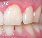
Colour
The normal gingival colour is coral pink and is produced by the tissue vascularity and modified by the overlying epithelium. Increase in vascularization or less epithelial keratinization will cause the gingiva to be more red. Reduction in vascularity (eg. fibrosis of the corium) or increase in epithelial keratinization will cause the gingiva to be more pale.
Consistency
Normal gingiva is firm and resilient in consistency. Destructive changes will cause the gingiva to be more edematous and reparative changes will cause the gingiva to be more fibrotic
Surface Texture
Loss of surface stippling is an early sign of gingivitis, but the absence of stippling does not mean absence of health.
Gingival Contour
Gingival enlargement
Stillman's clefts
McCall festoons